Showing all 2 results


Fluorescence microscopy differs from other microscopy methods in a variety of ways. Normal brightfield microscopy in a high power microscope takes light and passes it through a thin specimen on a glass microscope slide. Phase contrast microscopy begins with normal light, then shifts it out of phase and this phase shift is translated into a difference in viewing intensity. Darkfield microscopy is an illumination technique where the biological specimen is illuminated from the sides instead of from the bottom.
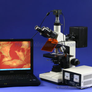
The technique used in fluorescence microscopy is far more different from all other technique because the light that can be seen under this kind of microscope is not the same light that was produced by the illumination source. The light that is viewed under this kind of microscope is really the light that has been produced by the fluorescence of the specimen under the microscope. Fluorescence microscopes utilizes illumination source that is capable of producing high intensity light. The high intensity light is made to go through the dichroic filter cube that encloses a fluorescence bandpass excitation filter. The dichroic filter cube permits only particular light wavelength to pass though it and arrive at the specimen under the microscope. Once the light that has passed through the dichroic filter comes in contact with the specimen, there is no further use for it and any amount of light that has been reflected back, either into the objectives of the microscope or on the dichroic filter, is again filtered out by the use of emission filter. When the specimen under the microscope starts to fluoresce, it emits a fluorescing light that goes through the fluorescence emission filter and it is then passed into the eyepieces of the microscope, producing the fluorescence image of the sample.
The selection of these fluorescence excitation filters and fluorescence emission filters is critical to achieving the proper fluorescence image. Different fluorescence specimens fluoresce at various wavelengths of light. Our fluorescence microscopes come with several dichroic filters of various wavelengths. If you need specialized custom made fluorescence filters, then we can provide that as well. We work with two of the best names in fluorescence microscopy, Chroma and Omega. Filters from these prestigious fluorescence filter companies will outperform standard fluorescence filters because they are custom made to your precise wavelength specifications and because they pass less undesired wavelengths. It is recommended to review your fluorescing biological sample and the fluorescing stain. For example, if you are using GFP, green fluorescence protein, that has a specific narrow light wavelength spectrum in which it will optically fluoresce. Your fluorescence filters should match this wavelength spectrum for optimal imaging. We are happy to work with you to ensure your fluorescence stain is matched to the correct fluorescence filters. If our stock filters do not match the needed wavelength properly, we can assist you to obtain custom made filters for optimal imaging quality.
If you are looking for a fluorescence microscope, please contact our technical sales staff who will be happy to assist you. Our epi-fluorescence microscopes are sold at a discount to pass the savings to our customers. Call today to lock in your deal on good quality fluorescence microscopy equipment.